Preeclampsia remains one of the leading causes of maternal and perinatal death worldwide, affecting approximately 2–7% of pregnancies.1
It is a condition that can develop suddenly and progress rapidly, leaving patients and clinicians with limited time to respond. Traditionally, preeclampsia was understood as a placental disorder, but recent insights point to a more complex origin. Research by Prof. Poon and Prof. Nicolaides (King's College Hospital, London) suggests that preeclampsia may in fact be a cardiovascular condition, explaining why women who experience it during pregnancy face a higher lifetime risk of heart failure and stroke.2
From Late Diagnosis to Early Prediction
Historically, preeclampsia was often diagnosed only after symptoms such as high blood pressure or swelling appeared.² Today, advances in ultrasound and biomarker-based screening are transforming how clinicians approach this disease.
Professor Liona Poon, developed an early risk-assessment model performed between 11 and 13 weeks of gestation.3
Her approach combines maternal risk factors, uterine-artery Doppler measurements (Pulsatility Index), mean arterial pressure (MAP) and placental growth factor (PlGF).³ According to the ASPRE trial, this combined model can identify approximately 75 percent of preterm preeclampsia cases with a 10 percent false-positive rate.4 Such predictive capability allows for timely intervention and potential preventive treatments.5
Ultrasound Technology and Precision Tools
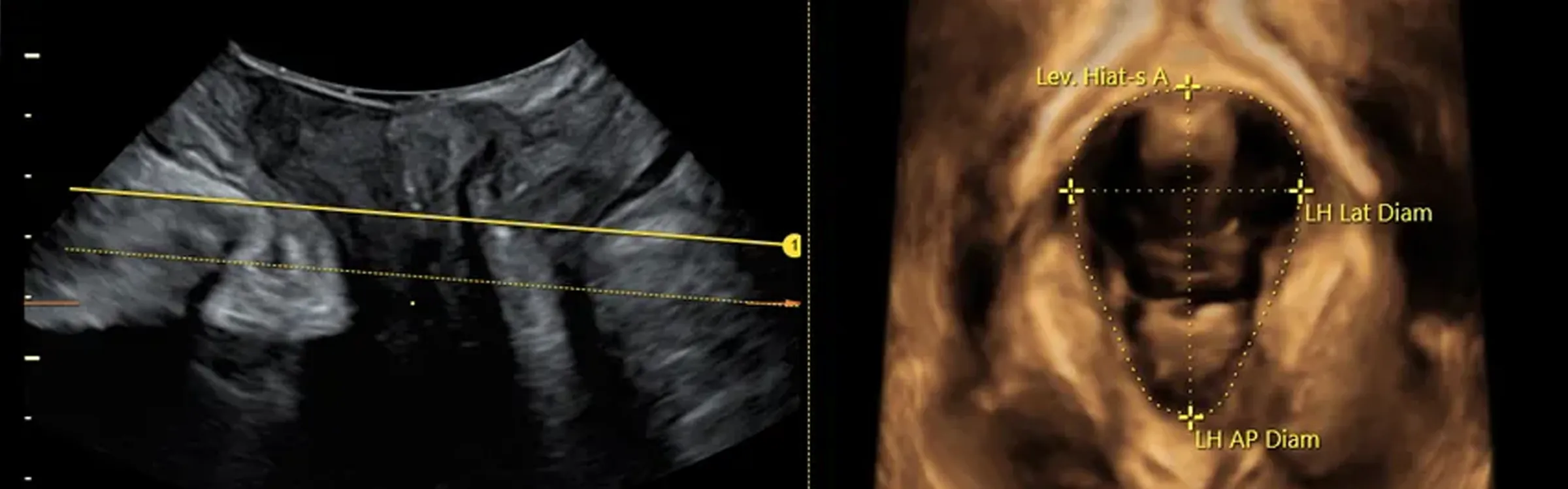
Accurate imaging is essential to effective screening, and this is where GE HealthCare’s Voluson™ ultrasound platform excels.3
Voluson systems offer advanced Doppler and visualization tools designed for detailed vascular assessment.3
- SlowflowHD enables clinicians to visualize low-velocity blood flow and placental microvascular perfusion, making even the smallest vessels visible.3
- Radiantflow provides fast, high-resolution imaging of tiny vessels, supporting precise uterine-artery evaluation.3
- HDlive™ technology uses customizable virtual lighting to enhance depth perception and reveal internal anatomical structures in unprecedented detail.3
These innovations make it easier to locate uterine arteries and accurately measure the pulsatility index, one of the key ultrasound biomarkers for preeclampsia.3
Integrating Data for Smarter Care
To streamline workflow, GE HealthCare developed ViewPoint™ 6, an intuitive software platform that integrates patient history, ultrasound measurements, and laboratory data into a single reporting environment.
Clinicians can input all relevant information — such as crown-rump length, uterine-artery PI, MAP, and PlGF — to calculate individualized preeclampsia risk directly within the program.³ “Many clinicians around the world are using ViewPoint to capture data. I can enter the data of uterine artery pulsatility index, mean arterial pressure, placental growth factor, and history — and calculate the risk, all in one program,” explains Prof. Liona Poon.³ This level of integration simplifies early screening and enhances the clinician’s ability to act proactively rather than reactively.3
A New Perspective on Maternal Health
Research continues to refine our understanding of preeclampsia.2
Prof. Nicolaides‘ and Prof. Poons‘ work underscores that there may be two distinct types — early-onset, driven by placental dysfunction, and late-onset, associated with maternal cardiovascular maladaptation.⁵ Recognizing these differences allows for more tailored monitoring and highlights the importance of ongoing cardiac assessment even after delivery.2
Conclusion
Ultrasound has become a cornerstone in the early detection and management of preeclampsia, offering clinicians powerful tools to assess both maternal and fetal health.3 By combining innovative imaging technologies such as SlowflowHD, Radiantflow, with integrated software solutions like ViewPoint™ 6, GE HealthCare is helping clinicians move from late diagnosis to true early prediction and prevention.3 This shift represents not only a technological evolution but a new standard in maternal care — one that could significantly reduce the global burden of preeclampsia.1,5
References
- World Health Organization. Make Every Mother and Child Count.Geneva: World Health Report; 2005.
2. Chaney P. A New Perspective on Preeclampsia and Maternal Death.GE HealthCare Women’s Health, Feb 2024. Access: https://www.volusonclub.net/empowered-womens-health/a-new-perspective-on-preeclampsia-and-maternal-death/ (last access: Oct 2025).
3. GE HealthCare Women’s Health. The Role of Ultrasound in Screening for Preeclampsia. 2024. Access: https://www.volusonclub.net/empowered-womens-health/case/preeclampsia-screening-ultrasound/ (last access: Oct 2025).
4. Rolnik D, Wright D, Poon L. ASPRE Trial: Performance of Screening for Preterm Preeclampsia. Ultrasound Obstet Gynecol. 2017; 50: 492–495.
5. O’Gorman N, Nicolaides K, Poon L. The Use of Ultrasound and Other Markers for Early Detection of Preeclampsia.Women’s Health (Lond) 2016; 12(2): 199–207.
Other Topics

AI in OB/GYN Ultrasound
The Pelvic Floor: More than “Just a Muscle”

Ultrasound in ART: A pivotal tool for infertility diagnosis and treatment success

The Role of Advanced Ultrasound Features in Infertility

The Emerging Impact of Handheld Ultrasound Across Obstetrics

Understanding Endometriosis – What, Why and How
Fetal Heart