
 Clinical specialty
The Evolving Role of Ultrasound in Labor
Intrapartum ultrasound is transforming labor and delivery by providing real-time, objective insights into labor progress, reducing the uncertainty associated with traditional digital vaginal examination (DVE), which is subjective and operator-dependent.1,2 Transperineal ultrasound offers reproducible, imaging-based assessment that improves clinical confidence and decision-making.1,2
Modern systems such as GE HealthCare Voluson™ platforms, including applications like Voluson™ SonoL&D, further enhance clinical practice through structured workflows, standardized measurements, and improved documentation. SonoL&D complements manual examination using 2D imaging and semi-automated measurements to evaluate fetal head position and descent during the second stage of labor, while also supporting patient communication through intuitive video and graphic visualization.3
Key benefits include:
- Objective assessment of cervical dilation, fetal head position, station, and descent1-4
- Higher accuracy and interobserver agreement than DVE1,2
- Reduced uncertainty and improved clinical confidence1,2
- Real-time visualization of fetal and maternal anatomy1-4
- Improved maternal comfort and reduced infection risk1,2
Ultrasound also enhances workflow and communication:
- Clear documentation and shared understanding among clinicians2,5
- Support for supervision and decision-making in busy units5
- Dynamic monitoring of labor progression using measurable parameters1-4
- Assessment of caput, skull molding, and fetal head rotation1-4
- Reliable assessment despite maternal obesity, fetal caput, or examiner variability, where digital examination may be inaccurate or limited1,6
- Improved efficiency and consistency in care2,5
Monitoring Labor Progress with Ultrasound
Transperineal ultrasound enables dynamic, repeatable assessment of fetal descent and cervical changes, providing objective insight into labor biomechanics.1,2,8 Clinicians can efficiently obtain key measurements—such as the angle of progression (AoP), head–perineum distance (HPD), and midline angle (MA)—to support decision-making during active labor.
Common parameters include:
• Fetal descent (AoP, HPD)1-4,6-8
• Sonographic cervical dilation (SCD)1-3
• Caput formation and skull adaptation1,2,5
• Fetal head position, rotation, and alignment1-3
Angle of Progression (AoP)
The AoP is a well-established parameter for assessing fetal head descent and predicting delivery outcomes.7 It is measured in the sagittal plane between the axis of the pubic symphysis and a line tangential to the fetal skull.4,7 Larger angles indicate greater descent and are associated with successful vaginal or operative delivery.7 Serial measurements allow reliable tracking of labor progression and are more accurate than digital examination when interpreted alongside other parameters.1-4
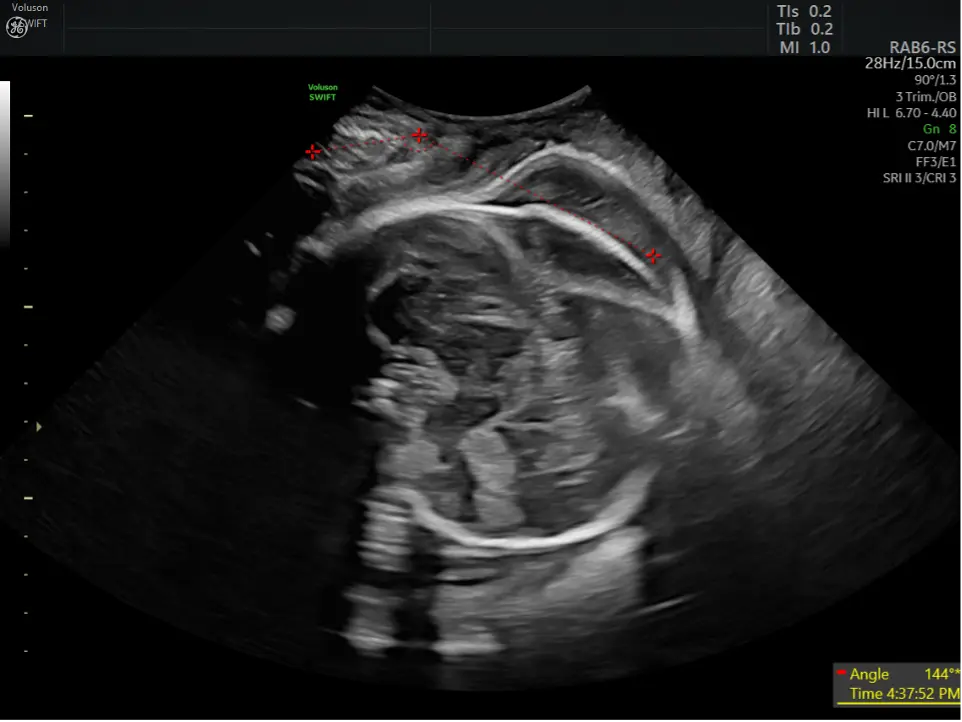
SonoL&D assisted documentation of Angle of Progression
Head–Perineum Distance (HPD) and Cervical Assessment
HPD measures the shortest distance between the fetal skull and perineum and decreases as labor progresses.1,2,8 Repeated measurements provide an objective method to assess descent, especially when caput limits clinical examination.2,8
Ultrasound also enables estimation of cervical dilation (SCD), offering a less invasive and more objective alternative to manual assessment.1,2
Fetal Skull Adaptation and Rotation
Transperineal ultrasound allows visualization of caput succedaneum and fetal skull molding, improving understanding of labor mechanics. It also enables assessment of fetal head rotation via parameters such as the midline angle, which is essential for safe operative delivery. 2,5
Additional signs, such as the “head up sign” and perineal muscle activity, can further inform labor progress and clinical decisions.2,5
Practical Considerations
For optimal imaging, the maternal bladder should be empty.1 Visualization may be limited when the fetal head is high, and gentle probe pressure is required to ensure image quality while maintaining patient comfort.1 Intrapartum ultrasound is particularly valuable in prolonged labor or when repeated assessment is needed.2
Clinical Value and Future Directions
Ultrasound enhances safety in operative vaginal delivery by confirming fetal head position and guiding correct forceps placement in real time.1,2,9 It also improves patient communication, as visual imaging and graphics help patients better understand labor progress and participate in decision-making.1
Current guidelines, including those from the Royal College of Obstetricians and Gynaecologists, recommend ultrasound assessment when fetal head position is uncertain, reinforcing its clinical value.1,9
Future developments include:
- AI-assisted measurements to improve reproducibility2
- Sonopartograms for visualizing labor progression2
- Predictive models combining ultrasound and clinical data2
- Expanded applications in pelvimetry and biomechanics2
Ultimately, intrapartum ultrasound improves accuracy, safety, and communication in labor management, particularly during the second stage, enabling more informed clinical decision-making.1,2 It enables continuous, data-driven monitoring of labor and supports more personalized care.1,2
References
- Ghi T, Eggebø TM, Lees C, et al. ISUOG Practice Guidelines: intrapartum ultrasound. Ultrasound in Obstetrics & Gynecology. 2018.
- Ramirez Zegarra R, Lizarraga Cepeda E, Ghi T. Intrapartum ultrasound. Best Pract Res Clin Obstet Gynaecol. 2025;101:102617.
- https://womens-health.net/clinical-specialty/labor-delivery/sonold-benefits
- Taychaburapanone P, Pongchaikaikiti P, Onraksa W, Chindasombatcharoen S, Suntipap M. Transperineal ultrasonography angle of progression measurement as a predictor of successful labor in pregnancy with induction of labor: a systematic review and meta-analysis. J Matern Fetal Neonatal Med. 2025;38(1):2537231.
- Malvasi A, Tinelli A. Intrapartum Ultrasonography for Labor Management. Springer. 2016.
- Malvasi A, Baldini GM, Difonzo T, Cerbone M, Cara I, Demarco M, Mappa I, Rizzo G, Vimercati A, Dellino M, et al. Intrapartum Ultrasound in Vacuum Operative Delivery: A Comprehensive Review and Proposal of the Novel Ultrasound Flexion Point Method. Diagnostics. 2026; 16(6):946.
- Barbera AF, Pombar X, Perugino G, Lezotte DC, Hobbins JC. A new method to assess fetal head descent in labor with transperineal ultrasound: the angle of progression. American Journal of Obstetrics and Gynecology. 2009.
- Eggebø TM, Gjessing LK, Heien C, et al. Prediction of labor and delivery by transperineal ultrasound in the second stage of labor. Ultrasound in Obstetrics & Gynecology. 2008.
- Royal College of Obstetricians and Gynaecologists. Operative Vaginal Delivery. Green-top Guideline No. 26. Updated 2020. RCOG; 2020.
JB01939AFG
Other Topics

The Role of Ultrasound in the Early Diagnosis and Follow-Up of Preeclampsia

AI in OB/GYN Ultrasound
The Pelvic Floor: More than “Just a Muscle”

Ultrasound in ART: A pivotal tool for infertility diagnosis and treatment success

The Role of Advanced Ultrasound Features in Infertility

The Emerging Impact of Handheld Ultrasound Across Obstetrics

Understanding Endometriosis – What, Why and How

Fetal Heart